A small (but glorious) world: The best microscope images of 2012
Ars Technica » Scientific Method 2012-10-24
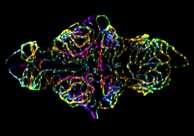
Most people know Nikon as a purveyor of pro and consumer-grade digital cameras. But the company's expertise with optics bleeds over into related markets—it's one of the science community's major suppliers of microscopes. And each year the company asks the community to send it some of their favorite images of tiny objects. A panel of scientists and journalists have chosen the best of this past year's submissions, which Nikon has placed on its Small World site.
We've gone through and picked out some of our favorite images from this year, and Nikon provided some high-resolution versions. In keeping with the Ars tradition, where possible, we'll tell you a bit more about the subjects than you might get from the brief description on the original site.
The grand prize winner at top highlights the blood vessels as they form in the brain of a zebrafish. The fish itself is transparent at this stage of development, and the blood vessels are tagged with a fluorescent protein, which allowed the researchers to image these tiny vessels at 20 times their normal size. The image is actually a composite of many individual images taken with a confocal microscope.
Read 11 remaining paragraphs | Comments